Tempo

Dose Optimized STEM

Tempo improves the amount of information obtained for a given electron dose, or the “information efficiency”, of STEM experiments. The concept is simple: There is a large diminishing return on information as more electrons are detected from each point in a STEM scan, so for each dwell period, quickly turn off the electron beam after a given number of electrons are counted (typically 1 – 25) and turn it back on at the start of the next dwell period. In this new imaging paradigm, TempoSTEM, pixel intensity is defined by the time taken to detect a fixed number of electrons, as opposed to the number of electrons detected in a fixed time. While this distinction may seem small the implications for minimizing specimen damage are substantial.

Tempo unit

Features at a Glance

Dose Optimization

Maximum information efficiency extracted from a minimum delivered electron dose

Real Time Imaging

Tempo outputs directly interpretable image signals that are compatible with most STEM controllers so no post processing is required

User Defined Trigger Thresholding

Flexibly balance dose or precision across full duty-cycle range by setting the blanking trigger condition

Detector Compatibility

Compatible with a wide variety of existing analog STEM detectors

Ease of Integration

A simple upgrade for any instrument with EDM Basic or EDM Synchrony

Specification

Quantity
Channels2 in, 2 out
Tempo voltage3.3 V or 5 V
Tempo resolution8 ns
Pixel clock input3.3V CMOS - 5V TTL
Blank signal output3.3V CMOS - 5V TTL
Signal rangeMax. ±10 V
Signal resolution14-bit
Sample rate125 Msps (per channel)
Tempo system diagram
Applicable Models

Signal to Noise Ratio vs Event Threshold Setting

TempoSTEM images of human macrophage cells recorded with the beam blanker triggering after various event numbers. Contrast represents scattering-rate values. Sample credit: Alexandra Porter (ICL) and Karin Muller (U. Cambridge).

Damage Reduction

Inital State
TempoSTEM image
After TempoSTEM imaging
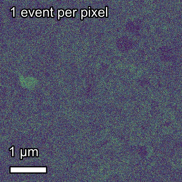
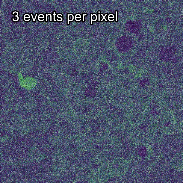
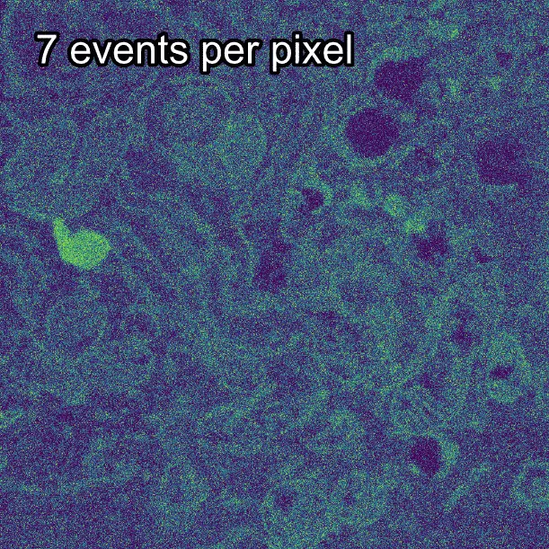
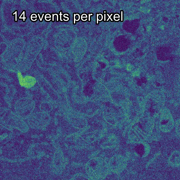
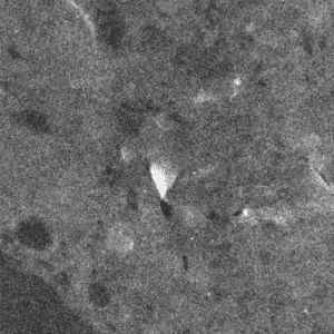
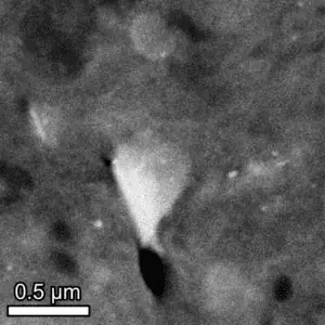
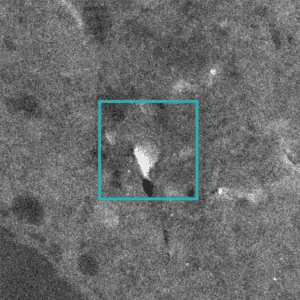
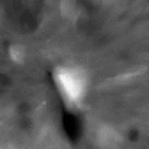
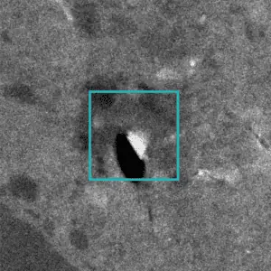
Example of reduced damage to a human macrophage sample when using TempoSTEM to image versus conventional STEM. After imaging with TempoSTEM (using 3 electrons per pixel, 10 averaged frames) the initial state is preserved. During and after conventional imaging (10 averages frames) the sample displays significant damage and distortions.
Conventional image
After conventional imaging
Sample credit: Alexandra Porter (ICL) and Karin Muller (U. Cambridge)

Equivalent Contrast

TempoSTEM Image
Conventional Image

Comparison of the image contrast between TempoSTEM and conventional STEM imaging of SrTiO3. Both images are shown in units of events per microsecond. The quantitative information offered by TempoSTEM is identical to conventional approaches. TempoSTEM image shows 210 electrons per pixel, conventional image uses approximately equivalent total average dose.